
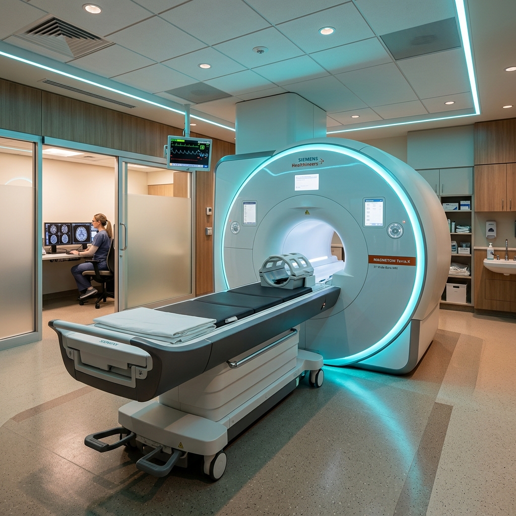
Magnetic Resonance Imaging (MRI) is one of the most powerful and non-invasive diagnostic tools in modern medicine. Every day, thousands of patients lie inside a large, cylindrical scanner, hear loud thumping noises, and emerge with highly detailed 3D cross-sections of their internal organs, brain tissues, and joints. But how did this technology come to be, and what makes it so unique?
The History: Who Invented the MRI?
The science behind MRI traces back to the mid-20th century. In 1946, researchers Felix Bloch and Edward Purcell discovered Nuclear Magnetic Resonance (NMR) in liquids and solids, earning them the Nobel Prize in Physics in 1952. For decades, NMR was used primarily as a chemical tool to evaluate molecule structure.
The breakthrough that turned NMR into a medical scanning machine came in the early 1970s. Dr. Raymond Damadian, an American physician and scientist, discovered that cancer tissues and healthy tissues responded differently to magnetic resonance signals. In 1977, Dr. Damadian and his team successfully conducted the very first human MRI scan using a home-built scanner named “Indomitable.” That first scan of a colleague’s chest took nearly five hours to generate a single image!
Simultaneously, scientists Paul Lauterbur and Peter Mansfield developed the mathematical equations and gradients necessary to turn signals into clear, two-dimensional images rapidly. They were awarded the Nobel Prize in Physiology or Medicine in 2003 for their pivotal advancements.
How Does it Work? (The Physics in Simple Terms)
Unlike X-rays or CT scans, an MRI scan uses zero ionizing radiation. Instead, it utilizes two natural components: a powerful magnetic field and radiofrequency waves. Here is the step-by-step process:
- Alignment: The human body is mostly water, which contains hydrogen atoms (protons). Normally, these protons spin randomly. When you enter the MRI’s strong magnetic field, these protons align themselves in the direction of the field (like microscopic compass needles).
- Excitation: The scanner emits a burst of radio waves, which temporarily knocks the protons out of alignment.
- Relaxation: When the radio waves stop, the protons snap back into alignment. As they do, they release energy in the form of radio signals.
- Imaging: Different tissues (fat, muscle, tumor, bone) snap back at different speeds, releasing different signals. Highly sensitive coils in the scanner capture these signals, and a computer converts them into the high-contrast 3D images your radiologist reads.
Fascinating MRI Facts You Might Not Know
- Incredible Magnet Strength: The strength of an MRI magnet is measured in Tesla (T). Standard medical MRI scanners operate at 1.5T to 3.0T. To put this in perspective, a 1.5T magnet is roughly 30,000 times stronger than the magnetic field of the Earth!
- Why is it so loud? The thumping and knocking noises you hear during a scan are caused by electric currents passing through the scanner’s gradient coils. The strong magnetic forces make the coils vibrate, producing sound. Earplugs or headphones are always provided to minimize the noise.
- Metallic Restrictions: Because the magnet is active 24/7 (even when not scanning!), metallic objects (like keys, phones, jewelry, or pacemakers) are strictly forbidden in the scan room. They can turn into dangerous projectiles or become deactivated.
Today, MRI scans are the gold standard for scanning the brain, spinal cord, ligaments, and organs, helping physicians diagnose neurological conditions, sports injuries, and cardiac anomalies with absolute precision.