
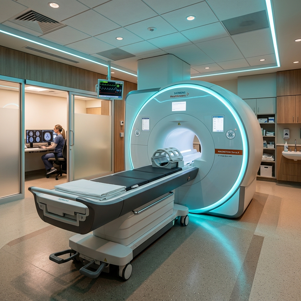
For nearly eighty years after Wilhelm Röntgen discovered X-rays in 1895, medical imaging was completely flat. Traditional X-rays compress 3D structures into a single 2D sheet, meaning overlapping bones and dense organs cast shadows on one another, occasionally hiding critical tissue damage. That changed in 1972 with the invention of Computed Tomography (CT), which sliced the human body digitally to give doctors a three-dimensional view inside.
The Beatles Connection: Music Profits Funding Medical Science
The invention of the CT scan is tied to one of the most famous bands in rock history: The Beatles.
In the 1960s, Godfrey Hounsfield, a brilliant British electrical engineer, was working at the research labs of EMI (Electric and Musical Industries). At the time, EMI was primarily an industrial conglomerate that happened to own Parlophone Records—the label representing The Beatles.
As Beatlemania took over the world, EMI accumulated unprecedented profits. Seeking areas to invest their capital, the company allowed Hounsfield to pursue a highly speculative and expensive project: using computers to combine multiple X-ray readings into clear 3D images. Thanks to the money generated by songs like “Help!” and “Yesterday,” Hounsfield successfully built the first commercial scanner. In 1979, Hounsfield and physicist Allan Cormack were awarded the Nobel Prize in Physiology or Medicine for their creation.
How Does it Work? (The Physics in Simple Terms)
A CT scan uses X-rays, but instead of taking a single shot, it captures thousands of exposures in a circular sweep:
- The Gantry Circle: The patient lies on a flat table that slides slowly into a large donut-shaped machine called the gantry.
- 360-Degree Rotation: Inside the gantry, an X-ray tube rotates rapidly around the patient. Opposite the tube is an array of electronic detectors.
- Cross-Section Slicing: As the scanner spins, it shoots narrow beams of X-rays through the body. The detectors record exactly how much radiation is absorbed by the different densities of bones, organs, and fluid.
- Computer Stacking: A powerful processing computer runs complex reconstruction algorithms to combine these raw rotational readings into hundreds of horizontal cross-sections (“slices”). These slices can be stacked together to render rotating 3D models of blood vessels, organs, and bones.
The Power of CT in Emergency Care
In trauma medicine, speed is the difference between life and death. While MRI scans deliver unparalleled detail for soft tissues, they can take 30 to 45 minutes to perform. A CT scan, on the other hand, can scan the entire chest, head, or abdomen in under 10 seconds.
This extreme speed makes CT the absolute gold standard for:
- Trauma Cases: Instantly detecting internal organ lacerations, pelvic fractures, and active hemorrhaging.
- Stroke Evaluation: Rapidly identifying whether a stroke is ischemic (blocked artery) or hemorrhagic (bleeding vessel) so the correct treatment can begin immediately.
- Lung & Chest Scans: Detecting embolisms, infections, and tumors with high resolution.
Fascinating CT Scan Facts You Might Not Know
- The First Brain Scan: The first patient ever scanned by a CT machine was in October 1971 at Atkinson Morley Hospital in London. The scan successfully located a dark, plum-sized cyst in the patient’s frontal lobe, proving the clinical capability of Hounsfield’s invention.
- Hounsfield Units (HU): Density in CT imaging is measured on a scale named in honor of Godfrey Hounsfield. Air is defined as -1,000 HU (appears pitch black), pure water is 0 HU, and dense bone registers between +1,000 and +3,000 HU (appears solid bright white).
- Blink of an Eye: Hounsfield’s first laboratory prototype required over nine days of data collection to compile a single slice. Today’s multi-slice CT scanners can capture 64, 128, or 256 slices in a fraction of a second, scanning a beating heart between heartbeats.
Today, CT scans remain one of the most critical imaging modalities, providing life-saving information in emergency settings and detail-rich guidance for cancer staging and cardiac evaluations.