
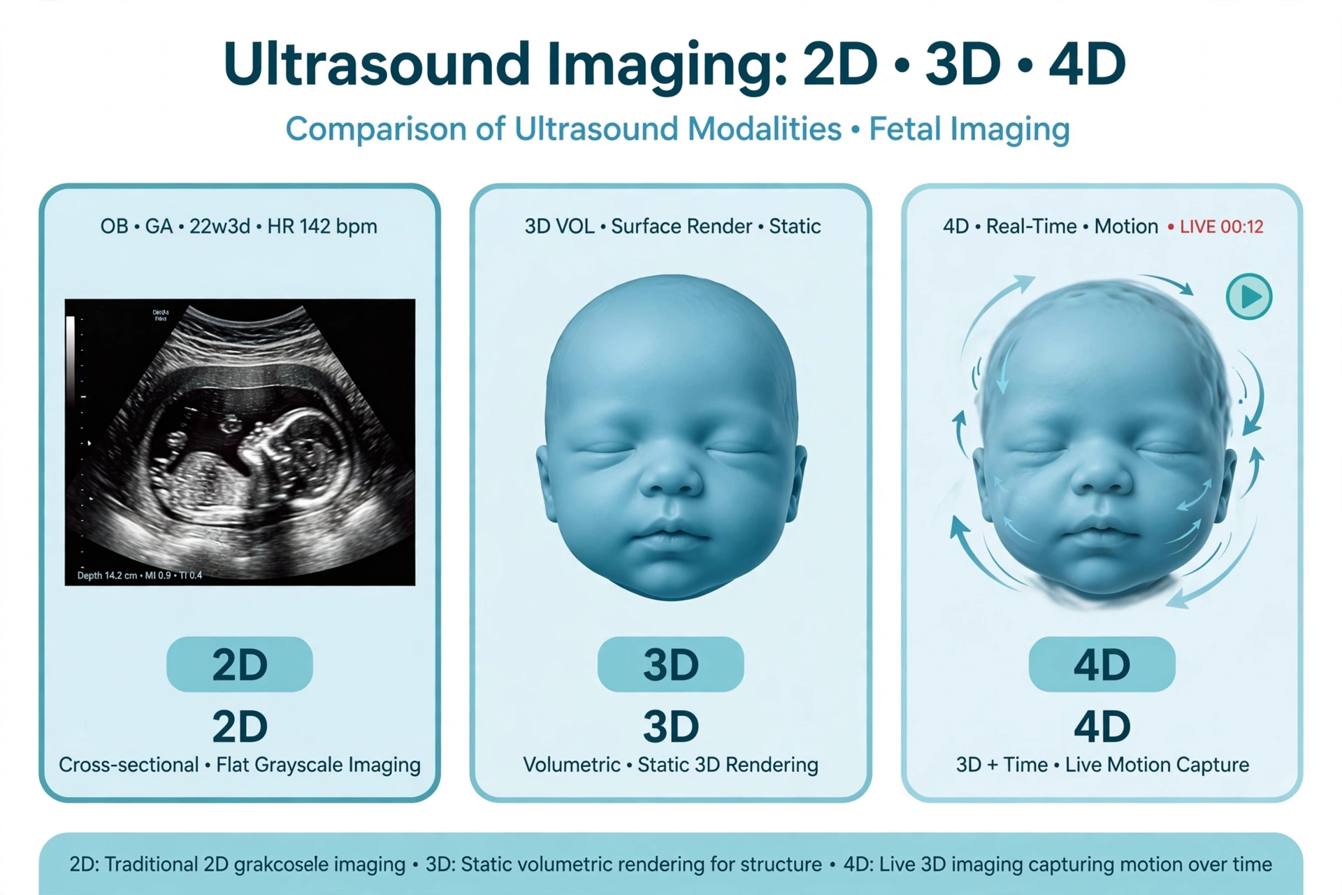
If your doctor has advised an ultrasound, you may have come across terms like 2D, 3D, and 4D. They all use the same safe sound-wave technology, but they present the images in different ways. Understanding the difference can help you know what to expect when you walk in for your scan.
What Is 2D Ultrasound?
2D ultrasound is the conventional, most widely used form of ultrasound imaging. It produces flat, two-dimensional images in shades of grey that show a cross-sectional “slice” of the inside of the body.
During a 2D scan, the sonologist moves the probe over the skin (or uses an internal probe for scans like TVS) and views the anatomy plane by plane. It has been the backbone of ultrasound diagnosis for decades, and it remains the primary tool for taking precise measurements — for example, measuring a baby’s head, abdomen, and thigh bone during pregnancy, or measuring the size of organs like the liver, kidneys, or thyroid.
Almost every diagnostic question in ultrasound is first answered with 2D imaging. It is reliable, fast, and sufficient for the vast majority of scans.
What Is 3D Ultrasound?
3D ultrasound builds on 2D technology. The machine acquires a volume of data — essentially many 2D slices captured in quick succession — and a computer reconstructs them into a three-dimensional image.
The most familiar example is the lifelike surface image of a baby’s face during pregnancy. 3D imaging can also display views that are difficult to obtain with 2D alone, such as the coronal plane of the uterus, which helps doctors assess the shape of the uterine cavity.
It is important to understand that 3D images are created from 2D data. The 3D view does not replace the standard 2D measurements; it adds an extra layer of visualisation that can be helpful in specific situations.
What Is 4D Ultrasound?
4D ultrasound is 3D ultrasound in real time. The fourth dimension is time — or motion. While a 3D scan gives a still three-dimensional image, a 4D scan shows that image moving live on the screen.
In pregnancy, this means parents can watch the baby move, yawn, or stretch in real time. From a medical perspective, 4D can help the doctor observe movement patterns and examine structures dynamically, though the core diagnostic measurements are still taken in 2D.
Key Differences at a Glance
- 2D ultrasound: Flat, cross-sectional grey images. The standard for measurements and routine diagnosis.
- 3D ultrasound: Still three-dimensional images reconstructed from 2D slices. Useful for surface views and certain anatomical planes.
- 4D ultrasound: Real-time moving 3D images. Shows motion as it happens.
- Technology base: All three use harmless high-frequency sound waves — none of them use radiation.
- Diagnostic role: 2D remains the workhorse. 3D and 4D are complementary tools that add visualisation in selected cases.
When Do Doctors Recommend Each Type?
The choice depends on the clinical question, and your doctor will advise what is appropriate for you. In general:
- Routine pregnancy scans, abdominal scans, pelvic scans, and thyroid scans are performed in 2D, which is all that is needed for accurate assessment and measurement.
- Detailed anomaly scans in pregnancy may use 3D/4D as an add-on to examine the baby’s face, spine, or limbs from additional angles.
- Certain gynaecological or cardiac evaluations may benefit from 3D reconstruction of specific structures.
No single type is “better” in every situation. A 3D/4D scan is not automatically more accurate than a 2D scan — accuracy depends on the skill of the sonologist, the quality of the equipment, and whether the right technique is used for the question being asked.
Is 3D/4D Ultrasound Safe?
Yes. 2D, 3D, and 4D ultrasounds all use the same sound-wave technology and do not involve ionising radiation. Ultrasound has been used in pregnancy and general medicine for decades and is considered safe when performed by trained professionals for a valid medical reason.
That said, medical bodies advise that ultrasound should be done when there is a clinical indication, not purely for entertainment. If you are offered a 3D/4D “keepsake” scan, it is worth discussing with your doctor whether it adds medical value in your case.
What to Expect During the Scan
A 3D/4D session is similar to a regular ultrasound. You will lie on an examination couch, gel will be applied to the skin, and the probe will be moved over the area being examined. A 3D/4D acquisition may take a little longer than a standard 2D scan because the machine needs to capture volume data.
For pregnancy scans, the clarity of 3D/4D images partly depends on the baby’s position, the amount of amniotic fluid, and the stage of pregnancy — so results can vary from person to person.
Talk to Your Doctor
If you are unsure which type of ultrasound you need, the simplest approach is to follow your doctor’s prescription. The referral usually specifies the scan required, and the imaging centre will use the appropriate technology. If you are curious about 3D/4D, ask your doctor whether it would add useful information in your particular case.
Medical Disclaimer
This article is for general information only and is not medical advice. Ultrasound technology and its applications should be discussed with your doctor, who can advise what is appropriate for your situation. If you have questions about a prescribed scan, please consult your doctor.
Book Your Ultrasound at Aura Care Centre
Aura Care Centre offers 2D, 3D/4D ultrasound and colour Doppler studies performed by experienced radiologists, with branches in Sector 38, Sector 15, and Sector 51, Gurugram. To book your scan, call +91 81301 01433 or message us on WhatsApp — we will help you schedule a slot at the branch most convenient for you.